Circulatory System in Animals
Senior 6 · Biology
Circulatory System in Animals
Lesson 4 of 5
25 min
If all the blood vessels of an adult were laid end to end, they would stretch for tens of thousands of kilometres, most of it capillaries finer than a hair. Blood leaves the heart at high pressure in thick-walled arteries, crawls through capillaries where it exchanges substances with the cells, and returns at low pressure in veins. On the way, some of its fluid leaks out to bathe the cells and must be returned. And if a vessel is cut, the blood must seal the hole within minutes. This lesson covers the vessels, the body fluids, clotting, and what goes wrong in cardiovascular disease, the leading cause of death worldwide.
Definition
Artery: a vessel that carries blood away from the heart, at high pressure. Arteriole: a small artery whose muscular wall controls blood flow into a capillary bed.
Capillary: the smallest vessel, with a wall one cell thick, where substances are exchanged between blood and tissue fluid.
Vein: a vessel that carries blood towards the heart, at low pressure; medium and large veins contain valves.
Tissue fluid: the fluid that bathes the cells, formed from plasma forced out of capillaries; it has almost no large proteins and no red blood cells. Lymph: tissue fluid that has drained into the lymphatic vessels.
Figure 1: Cross-sections of an artery, a vein and a capillary (not to the same scale).
| Artery | Vein | Capillary |
|---|---|---|
| Wall: Thick: endothelium, thick layer of smooth muscle and elastic fibres, outer collagen | Wall: Thin: same three layers, but little muscle and elastic tissue | Wall: One layer of endothelium only, often with gaps between cells |
| Lumen: Narrow, round | Lumen: Wide, often flattened | Lumen: About 7 to 10 µm, the width of one red blood cell |
| Pressure and flow: High, pulsing with each heartbeat | Pressure and flow: Low and steady | Pressure and flow: Falling; flow very slow |
| Valves: None, except at the base of the aorta and pulmonary artery | Valves: Yes, to stop backflow | Valves: None |
| How it suits its job: Elastic fibres stretch and recoil, smoothing the flow; muscle and collagen withstand high pressure; muscle in arterioles controls distribution | How it suits its job: Wide lumen offers little resistance; valves and the squeezing of nearby skeletal muscles keep blood moving towards the heart | How it suits its job: Thin wall gives a short diffusion distance; huge numbers give a huge surface area; slow flow gives time for exchange |
Blood is about 55 % plasma and 45 % cells by volume. Plasma is about 90 % water, carrying dissolved glucose, amino acids, salts, urea, hormones, carbon dioxide (mostly as hydrogencarbonate ions), and plasma proteins such as albumin, antibodies and fibrinogen. The cells are red blood cells (about 5 million per mm³; oxygen transport, Lesson 5), white blood cells (about 4 000 to 11 000 per mm³; defence) and platelets (cell fragments; clotting).
Tissue fluid forms because two forces act across the capillary wall (Figure 2):
Figure 2: Formation and return of tissue fluid, with typical values in kPa. Orange: fluid pushed out. Blue: water drawn back in.
At the arterial end, the push outwards wins, and water with small solutes (glucose, amino acids, oxygen, salts) is forced out to bathe the cells. At the venous end, the pull inwards wins, and water returns, bringing carbon dioxide and other wastes. About 90 % of the fluid returns this way. The rest drains into blind-ended lymph capillaries, flows through lymph nodes, where lymphocytes can meet any pathogens, and is returned to the blood in veins near the heart. If the balance fails, fluid collects in the tissues as oedema: for example, in kwashiorkor, a lack of dietary protein lowers the plasma proteins, so less water returns and the belly and legs swell.
Worked example 1: the pressures that form tissue fluid
The net filtration pressure is , where is hydrostatic pressure and oncotic pressure. Take , and (typical values). Calculate the net pressure at the arterial end () and at the venous end ().
When a vessel is damaged, three things happen quickly: the smooth muscle in the vessel wall contracts, platelets stick to the exposed collagen and to each other, forming a plug, and a cascade of enzyme reactions makes a clot.
Figure 3: The main steps of blood clotting. Dashed arrows: enzymes that catalyse the next step.
The cascade amplifies the signal: each activated enzyme molecule activates many molecules of the next, so a small injury produces enough fibrin within minutes. Clotting needs calcium ions and clotting factors made in the liver with the help of vitamin K. People with haemophilia inherit a faulty gene for one clotting factor, so their blood clots very slowly.
Atherosclerosis begins when the endothelium of an artery is damaged, for example by high blood pressure or the chemicals in tobacco smoke. White blood cells and low-density lipoprotein (LDL) cholesterol collect under the lining, forming a fatty atheroma (plaque), which later hardens with fibrous tissue and calcium. The lumen narrows and the wall stiffens.
Risk factors you cannot change: age, sex and family history. Risk factors you can: smoking, high blood pressure, high LDL cholesterol (diets high in saturated fat), diabetes, obesity, too much salt and alcohol, and physical inactivity.
In Rwanda, the 2022 national survey of non-communicable disease risk factors (Rwanda Biomedical Centre, STEPS survey) found raised blood pressure in 16.8 % of adults aged 18 to 69, and 52.1 % of adults had never had their blood pressure measured. Because hypertension causes no symptoms until it does damage, simply measuring it is one of the most effective ways to prevent heart attacks and strokes.

Worked example 2: why a small narrowing matters so much
For smooth flow through a tube, the flow rate at a given pressure is proportional to the fourth power of the radius (Poiseuille's law): . An atheroma reduces the radius of a coronary artery from to . Calculate the new flow as a percentage of the original, at the same pressure.
Limitation: real blood flow is pulsatile and blood is not a simple liquid, so the fourth-power rule is an approximation.

Warning
A suspected heart attack is an emergency. Signs include pain or pressure in the centre of the chest that may spread to the arm, jaw or back, breathlessness, sweating and nausea. Call for emergency medical help at once, and keep the person resting in a comfortable position. If they become unresponsive and are not breathing normally, start cardiopulmonary resuscitation (CPR) at once, pushing hard and fast in the centre of the chest (if you are not trained, give chest compressions only, as the emergency call-taker directs), and send for an automated external defibrillator if one is available.
Common mistakes. "Arteries carry oxygenated blood": arteries carry blood away from the heart (the pulmonary artery carries deoxygenated blood). "Tissue fluid is plasma": it lacks the large plasma proteins and red blood cells.
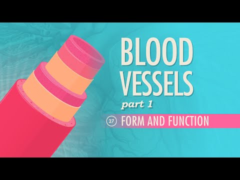
CrashCourse: "Blood Vessels, Part 1 - Form and Function" (Crash Course Anatomy & Physiology #27). Watch for the three layers of the vessel wall and how each type of vessel is built for its pressure.
Tip
Exam technique for tissue fluid. Always compare the two pressures at each end: "At the arterial end, hydrostatic pressure is greater than the oncotic pressure, so fluid is forced out; at the venous end, hydrostatic pressure has fallen below the oncotic pressure, so water moves back in by osmosis." Then mention the lymph for the excess.
Practice: vessels, body fluids, clotting and disease
Key points
Sources and credits
Figures 1 to 3, the tables, the worked examples and the practice questions are written by Ganzaa. The pressures in Figure 2 and Worked example 1 are typical textbook values.
This lesson adapts CC BY-SA material and is shared under CC BY-SA 4.0.