Gas Exchange in Animals and Plants
Senior 5 · Biology
Gas Exchange in Animals and Plants
Lesson 2 of 5
25 min
At a hospital clinic, a patient with a long-lasting cough is asked to take the deepest possible breath and then blow it all out, as hard and as long as possible, into a tube connected to a machine. The machine, a spirometer, measures how much air the lungs can move and how quickly. Doctors use it to diagnose asthma and chronic obstructive pulmonary disease, which you will meet in lesson 4. To read its results, you need to know how the lungs are built, how air is moved in and out, and what the different lung volumes mean.
Definition
Alveolus (plural alveoli): a tiny air sac at the end of a bronchiole, where gas exchange happens.
Tidal volume (TV): the volume of air breathed in, or out, in one breath at rest, about in an adult.
Vital capacity (VC): the largest volume of air that can be breathed out after the deepest possible breath in.
Residual volume (RV): the volume of air that is always left in the lungs, even after the most forceful breath out.
Pulmonary ventilation: the volume of air moved into the lungs in one minute.
Air passes from the nose and mouth through the trachea, which divides into two bronchi, one to each lung. These branch again and again into narrower bronchioles, which end in clusters of alveoli. Each lung contains hundreds of millions of alveoli, giving a total gas-exchange surface estimated at roughly (estimates range from about 50 to over 100 m²).
| Tissue | Trachea | Bronchi | Bronchioles | Alveoli | Function |
|---|---|---|---|---|---|
| Cartilage | C-shaped rings | Irregular plates | None | None | Holds the airway open when air pressure inside falls |
| Ciliated epithelium | Yes | Yes | Larger ones | None | Cilia beat to sweep mucus up to the throat |
| Goblet cells | Yes | Yes | Few | None | Secrete mucus that traps dust and microorganisms |
| Smooth muscle | Yes | Yes | Yes, relatively most | None | Contracts to narrow the airway; relaxes to widen it |
| Elastic fibres | Yes | Yes | Yes | Yes | Stretch on breathing in and recoil to help push air out |

Air moves from high to low pressure. The lungs have no muscles of their own; breathing works by changing the volume of the thorax, which changes the pressure inside it (for a fixed amount of gas, pressure falls when volume rises).
| Inspiration (breathing in) | Expiration at rest (breathing out) | |
|---|---|---|
| External intercostal muscles | Contract: ribs move up and out | Relax: ribs move down and in |
| Diaphragm | Contracts and flattens | Relaxes and domes upwards |
| Volume of thorax | Increases | Decreases |
| Pressure in lungs | Falls below atmospheric | Rises above atmospheric |
| Air movement | Into the lungs | Out of the lungs |
Quiet expiration is mostly passive: the muscles relax and the stretched elastic fibres recoil. In forced expiration, as when coughing or blowing into a spirometer, the internal intercostal muscles contract to pull the ribs down, and the abdominal muscles push the diaphragm up.
A simple spirometer is a chamber of oxygen floating on water. A person breathes in and out of it through a mouthpiece, wearing a nose clip, and the chamber rises and falls. A pen, or a sensor, records the movement as a trace (Figure 3). Breathed-out carbon dioxide is absorbed by soda lime in the circuit.
Two volumes add together to make a capacity: vital capacity , and total lung capacity . IRV and ERV are the inspiratory and expiratory reserve volumes: the extra air that can be breathed in, or out, beyond a normal breath. The residual volume cannot be measured with a spirometer, because it never leaves the lungs. Vital capacity is larger in taller people, in men than in women of the same height, and in trained athletes; it falls with age and with lung disease.
Worked example 1: pulmonary ventilation at rest and in exercise
At rest, a student's spirometer trace shows a tidal volume of and 4 breaths in 20 seconds. During exercise, the tidal volume is and the breathing rate is 30 breaths per minute. Calculate the pulmonary ventilation in each case, and the factor by which it rises.
Note: both deeper and faster breathing contribute, but here the tidal volume rose 4.4 times and the rate only 2.5 times.
Worked example 2: oxygen uptake from a spirometer trace
In a spirometer with soda lime, the troughs of a resting trace fall steadily by over 1.5 minutes. Explain why the trace drifts down, and calculate the rate of oxygen uptake in .
Check: without the soda lime, the volume would barely change, because about as much carbon dioxide would be breathed out as oxygen was taken in.
Warning
Common mistakes and safety
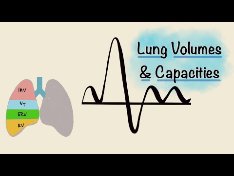
Byte Size Med: "Lung Volumes and Capacities | Spirogram". Watch for how each volume and capacity is read from the trace, and why the residual volume cannot be measured by spirometry; compare the definitions with Figure 3.
Tip
Memory hook: a capacity is always made of two or more volumes. Read a trace from the bottom up: RV, ERV, TV, IRV: "Rwanda's Exams Test Intelligence". VC skips the RV at the bottom; TLC includes everything.
Practice: human gas exchange and ventilation
Key points
Sources and credits
Figures 3 and 4, the tables, the worked examples and the practice questions are by Ganzaa. The volumes in Figure 3 are illustrative.
This lesson adapts CC BY-SA material and is shared under CC BY-SA 4.0.