Chemical Basis of Life
Senior 4 · Biology
Chemical Basis of Life
Lesson 5 of 5
25 min
Break an egg into a hot pan and the clear, runny egg white turns solid and white within a minute. Cool it down and it stays white. Egg white is mostly water and protein, and heat has changed the shape of the protein molecules for good, although not one of their amino acids has been removed. That tells you the key idea of this lesson: a protein works because of its precise three-dimensional shape, and that shape is decided by the order of its amino acids.
Proteins do almost every job in a cell: enzymes, antibodies, haemoglobin, insulin, the actin and myosin of muscle, the collagen of skin and bone, the keratin of hair. They are also the nutrient most linked with growth: the Rwanda Demographic and Health Survey 2019–20 found 33 % of children under five stunted (too short for their age), a sign of long-term undernutrition.
Definition
Amino acid: the monomer of proteins: a central carbon atom bonded to an amino group (), a carboxyl group (), a hydrogen atom and a variable R group. Proteins use 20 different amino acids. Essential amino acids (about nine for adults) cannot be made by the body and must be eaten.
Peptide bond: the C–N bond formed by condensation between the carboxyl group of one amino acid and the amino group of the next.
Polypeptide: a chain of many amino acids joined by peptide bonds. A protein is one or more polypeptides folded into a functional shape.
Denaturation: a change in the shape of a protein, caused for example by heat or extreme pH, that destroys its function without breaking its peptide bonds.
Prosthetic group: a non-protein part permanently attached to a protein, such as the haem of haemoglobin; such a protein is a conjugated protein.
Figure 1. Every amino acid has the same backbone; only the R group differs. A peptide bond forms by condensation and is broken by hydrolysis, as in digestion by protease enzymes.
Because one water molecule is lost for each peptide bond, a single chain of amino acids has peptide bonds. The R groups decide the properties of each amino acid: some are non-polar and hydrophobic (valine, leucine), some polar (serine), some charged (lysine is positive, glutamic acid negative), and cysteine contains sulfur.
Figure 2. The four levels of protein structure, simplified.
| Level | What it is | Bonds that hold it |
|---|---|---|
| Primary | The sequence of amino acids in the polypeptide, set by a gene | Peptide bonds (strong, covalent) |
| Secondary | Regular coiling of the backbone into an -helix, or folding into a -pleated sheet | Hydrogen bonds between the C=O of one peptide group and the N–H of another |
| Tertiary | The precise three-dimensional folding of the whole polypeptide | Between R groups: hydrogen bonds; ionic bonds between charged groups; disulfide bridges (S–S, covalent) between two cysteines; hydrophobic interactions between non-polar groups, which cluster inside away from water |
| Quaternary | Two or more polypeptides fitted together, sometimes with prosthetic groups | The same bonds as tertiary, between the chains |
Change the primary structure and the folding can change too. That is why a single wrong amino acid can wreck a protein, and why heat and pH, which break hydrogen and ionic bonds, denature it.
Worked example 1: counting peptide bonds in haemoglobin
Haemoglobin has four polypeptide chains: two chains of 141 amino acids and two chains of 146. (a) How many amino acids are there in one molecule? (b) How many peptide bonds, and how many water molecules were released in making them?

| Feature | Globular protein, e.g. haemoglobin | Fibrous protein, e.g. collagen |
|---|---|---|
| Shape | Folded into a compact, roughly spherical shape | Long, thin strands; three polypeptides wound into a triple helix, cross-linked into fibres |
| Solubility in water | Soluble: hydrophilic R groups on the outside | Insoluble |
| Amino acid sequence | Irregular | Repetitive (in collagen, every third amino acid is glycine, the smallest, which lets the chains pack tightly) |
| Role | Metabolic: transport (haemoglobin), enzymes, hormones (insulin), antibodies | Structural: strength in tendons, skin, bone and blood-vessel walls (collagen); hair and nails (keratin) |
Haemoglobin is a globular, conjugated protein. Each of its four chains holds one haem group, whose binds one molecule, so one haemoglobin molecule carries up to four .
Worked example 2: one amino acid, one disease
In sickle cell anaemia, the amino acid glutamic acid in the chain of haemoglobin is replaced by valine. Glutamic acid has a charged R group; valine's is non-polar. The change lies on the outside of the molecule. Explain why this one change makes red blood cells sickle-shaped when oxygen is low.

The depth of the purple colour increases with protein concentration, so, like Benedict's test, it can be made semi-quantitative with colour standards or a colorimeter. Sodium hydroxide is corrosive: wear eye protection.
Animal foods (milk, eggs, meat, fish) contain all the essential amino acids. Plant proteins are often short of one: beans are relatively low in methionine and cereals such as maize in lysine. Eating beans together with maize, sorghum or cassava-leaf dishes in the same day supplies a complete set, which is one reason why the traditional mixed Rwandan plate of beans and a starchy food works well.

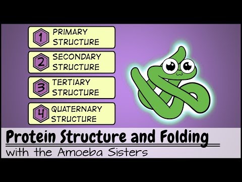
Amoeba Sisters: "Protein Structure and Folding". Watch for the four levels of structure (from about 2:06) and what denaturing does to a protein (from about 6:40).
Warning
Common mistakes
Tip
Memory hook for tertiary bonds: "HIDS": Hydrogen bonds, Ionic bonds, Disulfide bridges, hydrophobic interactions that Shield the core. In any question about a protein's function, connect: primary sequence → folding → specific shape → function (active site, binding site or strong fibre).
Practice: proteins
Key points
Further reading for this unit
Sources and credits
Quick check