Structure and Functions of the Human Gas Exchange System
Senior 1 · Biology
Structure and Functions of the Human Gas Exchange System
Lesson 3 of 5
25 min
Place both hands flat on your lower ribs, fingertips almost touching at the front, and take a slow, deep breath in. Your hands move up and apart. Now put one hand on your belly, just below the ribs, and breathe in again: it pushes outwards.
The lungs cannot have done this. Lung tissue contains no muscle that could make it expand; it is soft and elastic, like the goat's lungs in Lesson 1. So what makes air rush into your lungs 12 or more times a minute? This lesson answers with an experiment, a diagram and a model.
You need: a tape measure marked in centimetres (a tailor's tape is ideal), a partner, and a results table.
Safety. Breathe normally between readings: many deep breaths in a row can make you dizzy. If you have asthma, take readings slowly or act as the recorder instead.
Table 1: Sample results for Mugisha, aged 13 (sample data, for illustration)
| Reading | Chest after breathing out (cm) | Chest after breathing in (cm) |
|---|---|---|
| 1 | 71.0 | 76.5 |
| 2 | 70.5 | 77.0 |
| 3 | 71.5 | 76.0 |
Worked example 1: chest expansion from Table 1
Conclusion: Mugisha's chest gets about 5.5 cm bigger round the middle when he breathes in deeply. Taking the mean of three readings makes the result more reliable than a single reading.
Seal the tip of a plastic syringe with your finger and pull the plunger out. The space inside gets bigger, but no air can get in, so the air inside is spread more thinly: its pressure falls, and you feel the plunger being pulled back. Take your finger away and air rushes in. Air always flows from a place of higher pressure to a place of lower pressure.
Your chest works like that syringe. The lungs are sealed inside the thorax, and the pleural membranes make them follow the chest wall wherever it goes. So when muscles make the thorax bigger, the lungs are pulled open, the pressure inside them falls below the pressure of the air outside, and air flows in through the trachea.
Figure 1: Breathing in and breathing out at rest. Orange arrows: movement of the ribs and diaphragm. Blue arrows: air.
Table 2: Inhalation and exhalation compared
| Feature | Inhalation (breathing in) | Exhalation (breathing out, at rest) |
|---|---|---|
| Diaphragm | contracts and flattens, moving down | relaxes and returns to its dome shape, moving up |
| External intercostal muscles | contract | relax |
| Ribs and sternum | move up and out | move down and in |
| Volume of the thorax | increases | decreases |
| Pressure in the lungs | falls below the air pressure outside | rises above the air pressure outside |
| Air | flows in | flows out |
Breathing out at rest needs no effort. The muscles simply relax, and the stretched, elastic lungs spring back, like a stretched rubber band let go. Breathing out hard, as when you cough, shout or blow up a balloon, is different: the internal intercostal muscles contract to pull the ribs down and in, and the muscles of the belly push the diaphragm up harder. Some books give only this forced version, and say that the internal intercostal muscles contract in every breath out.
Definition
Inhalation (inspiration): breathing in. The thorax gets bigger, and air flows into the lungs.
Exhalation (expiration): breathing out. The thorax gets smaller, and air flows out of the lungs.
External intercostal muscles: the outer layer of muscles between the ribs. They contract to lift the ribs up and out when you breathe in.
Internal intercostal muscles: the inner layer of muscles between the ribs. They contract to pull the ribs down and in when you breathe out hard.
Air pressure: the push of the air on every surface it touches. When the same amount of air fills a bigger space, its pressure falls.
Warning
The lungs do not suck air in by themselves. They have no muscles to expand with. The diaphragm and intercostal muscles make the thorax bigger; the lungs are pulled open with it; the pressure inside falls; and only then does air flow in.
Watch the order of cause and effect. The volume changes first, then the pressure, and the air moves last. "The chest gets bigger because air comes in" has it backwards.
A contracting diaphragm moves down, not up. When its muscle contracts, the dome is pulled flatter and lower. It domes up again when it relaxes.
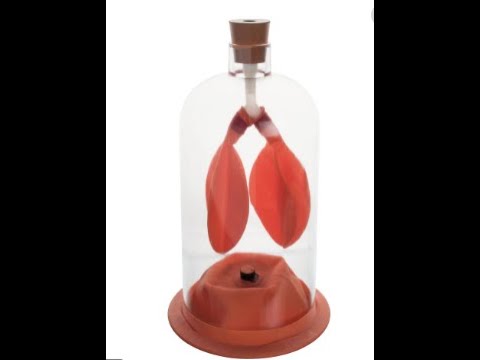
A bell jar is a glass jar with an open bottom. In the model, a Y-shaped glass tube passes through a bung in its top, with a balloon on each branch, and a rubber sheet is tied over the open bottom. Each part stands for a part of the body.
Figure 2: The bell-jar model. A: the sheet is pushed up. B: the sheet is pulled down, and the balloons inflate.
When the sheet is pulled down (B), the volume inside the jar increases, so the pressure in the jar falls. The air outside, pushing down the tube, is now at a higher pressure than the air in the jar around the balloons, so air rushes in and the balloons inflate, just as the lungs do when the diaphragm flattens. Push the sheet up (A) and the opposite happens: the balloons shrink and air leaves through the tube.
Make your own. Cut the bottom off a clear plastic bottle, push a drinking straw through a hole in the cap with a small balloon taped airtight to its end inside the bottle, and seal the gap round the straw with plasticine. Stretch a cut-open balloon over the cut end as the diaphragm. Pull its knot down and the small balloon inflates. Safety: your teacher should cut the bottle; cover the cut edge with tape, because it is sharp.
Worked example 2: how good is the bell-jar model?
Evaluate the bell-jar model as a model of breathing.
Judgement: the model is useful for explaining how the diaphragm causes the pressure changes that move air, but it leaves out the ribs and does not look like a real lung.
If you have a chest X-ray at a hospital, the radiographer will ask you to take a deep breath in and hold it. Now you can explain why: a deep breath flattens the diaphragm and lifts the ribs, so the lungs are as large as possible and more of them shows on the image, clear of the diaphragm and the heart.

Sean Clifford: "Bell jar model of the human breathing system". Watch the balloons as the rubber sheet moves, and name the part of the body that each piece of the model stands for.
Tip
Think of the syringe. Pull the plunger: the space gets bigger, the pressure falls, air rushes in. In your chest the plunger is the diaphragm, and the barrel is the rib cage. For exam answers, write the chain in order: muscles contract volume increases pressure falls air flows in.
Practice: the breathing mechanism
Key points
Sources and credits
Figures 1 and 2, the experiment, Tables 1 and 2, the worked examples and the practice questions are written by Ganzaa.