Radiation and Medical Imaging
Senior 6 · Physics
Radiation and Medical Imaging
Lesson 2 of 5
25 min
Every radiographer, nuclear-medicine technologist and radiotherapy physicist wears a small badge clipped to the chest. It does not beep or flash. Once a month it is posted to a laboratory, which heats or scans the material inside and sends back a single number in millisieverts. Over a working life those numbers are added up, and the law says the total must stay below fixed limits. To understand what the badge measures, you need three dose quantities, each answering a different question: how much energy was absorbed, how harmful that kind of radiation is, and how much the exposed organs matter to the whole person.
When ionising radiation passes through tissue, the secondary electrons and ions of Lesson 1 eventually turn the energy into chemical change and a little heat. The basic quantity is the energy deposited per unit mass:
Definition
Absorbed dose is the energy of ionising radiation absorbed per unit mass of material. Its SI unit is the gray (Gy): . Example: a tumour of mass 0.050 kg that absorbs 0.10 J has received .
Worked example 1: a dangerous dose is a tiny amount of heat
The World Health Organization gives the threshold for acute radiation syndrome as about 1 Sv; for gamma rays this is an absorbed dose of about 1 Gy. A person of mass 60 kg receives 1.0 Gy to the whole body. (a) How much energy is absorbed? (b) Treating the body as water (), by how much does its temperature rise?
Solution
(a) .
(b) .
Interpretation: 60 J is the energy of a cup of tea cooling by a fraction of a degree. The harm comes not from heat but from where the energy goes: into breaking bonds in DNA and other key molecules, as in Lesson 1.
The same absorbed dose does more biological damage when the radiation has a high LET, because the ionisation is packed so closely that DNA is broken in several places at once. The absorbed dose is therefore multiplied by a radiation weighting factor :
| Radiation | |
|---|---|
| photons (X-rays, gamma rays) of all energies | 1 |
| electrons (β⁻, β⁺) and muons | 1 |
| protons | 2 |
| alpha particles, fission fragments, heavy ions | 20 |
| neutrons | 2.5 to 20, depending on energy |
Radiation weighting factors recommended by the International Commission on Radiological Protection (ICRP Publication 103, 2007).
Definition
Equivalent dose in an organ or tissue is the absorbed dose multiplied by the radiation weighting factor, summed over the types of radiation: . Its unit is the sievert (Sv); has no unit, so too. Example: 1 mGy of alpha radiation in the lung gives .
Medical exposures are rarely uniform: a dental radiograph irradiates the jaw, a thyroid scan the neck. Organs also differ in how readily radiation induces cancer in them. Each equivalent dose is therefore multiplied by a tissue weighting factor , and the results are added:
| Tissue or organ | each |
|---|---|
| red bone marrow, colon, lung, stomach, breast, remaining tissues | 0.12 |
| gonads | 0.08 |
| bladder, oesophagus, liver, thyroid | 0.04 |
| bone surface, brain, salivary glands, skin | 0.01 |
Tissue weighting factors from ICRP Publication 103 (2007). Check: .
Because the factors add up to 1, a uniform whole-body equivalent dose of 2 mSv is an effective dose of 2 mSv. The effective dose is the number a personal dosimeter estimates and the number used to compare examinations.
Definition
Effective dose is the sum, over all irradiated organs, of each organ's equivalent dose multiplied by its tissue weighting factor. Unit: sievert (Sv). It is a protection quantity, designed for comparing exposures and setting limits for people in general, not a precise measure of one individual's risk.
Worked example 2: two partial-body exposures
(a) Radon gas is the main source of natural radiation (WHO). Over a year, the alpha particles from radon and its decay products give a person's lungs an absorbed dose of 0.50 mGy. Find the equivalent dose to the lungs and the effective dose. (b) A thyroid scan gives the thyroid an absorbed dose of 20 mGy from gamma rays, and other organs a negligible dose. Find the effective dose.
Solution
(a) . .
(b) . .
Comment: the thyroid in (b) received a far larger absorbed dose (20 mGy against 0.50 mGy), yet its effective dose is smaller, because gamma rays have and the thyroid is a smaller fraction of the whole-body risk.
Figure 2.1: The two kinds of biological effect. In (b) the straight line below about 100 mSv is an assumption used for protection, not a measurement.
| Source of exposure | Typical effective dose |
|---|---|
| natural background, world average (UNSCEAR) | 2.4 mSv per year |
| dental radiograph | 0.005 mSv |
| chest radiograph | 0.1 mSv |
| CT of the brain | 1.6 mSv |
| CT of the abdomen and pelvis | 7.7 mSv |
| whole-body PET/CT | 22.7 mSv |
Medical values for adults from RadiologyInfo.org (American College of Radiology and RSNA); individual doses vary with the machine and the patient. Ultrasound and MRI give no ionising dose at all.
A radionuclide given to a patient (Lesson 5) leaves the body in two ways at once: its nuclei decay, with the physical half-life of Unit 5, and the body excretes the substance (in urine, sweat and breath), with a biological half-life . Both are random processes with constant probabilities per unit time, so their rates add:
Since , dividing by gives the effective half-life:
Condition: biological removal must itself be exponential (a constant fraction excreted per unit time), which is a good first model. is always shorter than both and ; if one is much shorter than the other, is close to the shorter one.
Figure 2.2: Physical decay and biological removal together empty the body faster than either alone (biological half-life illustrative).
Worked example 3: technetium-99m in a patient
Technetium-99m has a physical half-life of 6.01 h. In a certain compound its biological half-life is 24 h. (a) Find the effective half-life. (b) What fraction of the injected activity remains in the patient after 24 h? (c) Compare with physical decay alone.
Solution
(a) .
(b) , about 3.1%.
(c) Physical decay alone: , about 6.3%. Excretion halves what remains after a day.
The international Basic Safety Standards (IAEA General Safety Requirements Part 3, 2014, co-sponsored by the WHO) apply three principles to every use of ionising radiation:
| Group (IAEA Basic Safety Standards) | Effective dose limit |
|---|---|
| radiation workers | 20 mSv per year averaged over 5 years, and not more than 50 mSv in any single year |
| members of the public | 1 mSv per year |
Workers' doses are measured with personal dosimeters. In a thermoluminescent dosimeter (TLD), radiation lifts electrons into traps in a crystal such as lithium fluoride; when the crystal is later heated, the electrons fall back and emit light whose total amount is proportional to the dose. Electronic personal dosimeters use a small semiconductor detector, show the dose at once and sound an alarm at a preset dose rate.


Worked example 4: weighing a risk against a benefit
A CT scan of the abdomen and pelvis gives an adult an effective dose of about 7.7 mSv. (a) How many years of average natural background (2.4 mSv per year) is this? (b) Using the LNT model and a nominal risk of 5% per sievert, estimate the extra lifetime chance of cancer. (c) Evaluate whether the scan should be done for a patient with suspected appendicitis.
Solution
(a) years.
(b) , roughly 1 in 2600.
(c) The estimated risk is small and is itself uncertain: it rests on the LNT assumption and is a population average, higher for children and lower for older adults. A missed, burst appendix is an immediate threat to life. The scan is justified if it will change the treatment, but ALARA still applies: in a child or a pregnant woman, ultrasound, which gives no ionising dose, would usually be tried first.
Warning
Common mistakes. (1) Treating gray and sievert as the same: they have the same base units, but the sievert includes the weighting factors, so 1 Gy of alpha radiation is 20 Sv. (2) Adding half-lives: the effective half-life is shorter than both, so it can never be . (3) Saying "below 100 mSv there is no risk": the risk is too small to measure, and protection assumes it is not zero. (4) Using the 5% per sievert figure to predict one person's fate: it is a population average for planning.
Tip
Climb the ladder of dose with three questions. Gray: how much energy per kilogram? Sievert (equivalent): how damaging is this radiation? () Sievert (effective): how much do these organs matter to the whole person? (, then add.) For half-lives, remember that rates add, so reciprocals add.
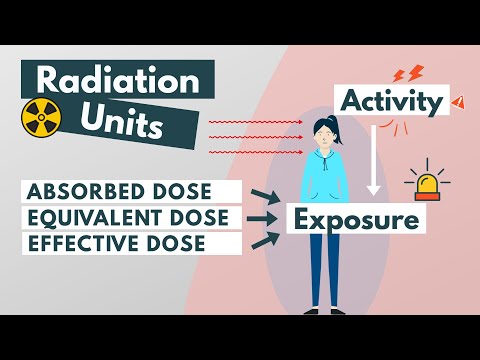
Radiation units: Absorbed, Equivalent & Effective dose, by Dr. Paulien Moyaert. Watch how the gray becomes a sievert through the two weighting factors, then check her factors against the tables in this lesson.
Practice
Key points
Sources and credits
The two diagrams, the worked examples and the practice exercises are written by Ganzaa.